Bones In Leg Diagram - Leg Anatomy / Joints of hand anterior view, lateral view, right hand.
Bones In Leg Diagram - Leg Anatomy / Joints of hand anterior view, lateral view, right hand.. The tarsal bones in the foot are located amongst tibia, metatarsal bones, and fibula. Each toe has three tiny bones, except for your big toe, which has just two. The smaller lateral bone of the lower leg. The diagram of bones in the ankle and foot is given below: Related posts of bones leg diagram picture bone anatomy of upper extremity.
The ankle is a bit different from the wrist; The patella (kneecap) is the sesamoid bone in front of the knee. This area is commonly referred to as the calf. Some types of leg pain can be traced to problems in your lower spine. It is where the lower leg bones connect to a large bone in the foot called the talus (say:
Leg Fracture What You Need To Know from www.drugs.com There are in all 7 bones, which fall under tarsal bones category. Most leg pain results from wear and tear, overuse, or injuries in joints or bones or in muscles, ligaments, tendons or other soft tissues. At the same time, the bones and joints of the leg and foot must be strong enough to support the body's weight while remaining. The ankle is a bit different from the wrist; The bones together make up the hip. This image is an edited version of this image that was created by user:ladyofhats (mariana ruiz villarreal). The head of the fibula. 10 october 2007 (original upload date) source:
The talocrual joint is made up of three main bones.
The knee joint is the largest joint in the body and is primarily a hinge joint, although some sliding and rotation occur. This area is commonly referred to as the calf. Disposition of rotator cuff muscles diagram. The hip itself is a ball and socket joint, much like the shoulder.the structures necessary to create this joint are the socket, the joint capsule, muscle, ligaments, and the neck. The pubis, ischium, and ilium together constitute the pelvis while the thigh bone is the femur. Many muscles that move the trunk and legs, such as our abdominal muscles, attach to the hip bones. At the same time, the bones and joints of the leg and foot must be strong enough to support the body's weight while remaining. Labeled human leg bones created for use in leg bone. The bones together make up the hip. The lower leg is comprised of two bones, the tibia and the smaller fibula. The lower limb contains 30 bones. Also called the shin bone, the tibia is the longer of the two bones in the. The patella (kneecap) is the sesamoid bone in front of the knee.
The human leg consists of 8 bones, 4 per leg. But the main part of the foot is similar to the hand, with five bones. The bones together make up the hip. Diagram and names of leg bones, diagram of foot and leg bones, diagram of leg bones, diagram of lower leg bones, diagram of the bones in your leg, bone, diagram and. With different grades of sprains depending on severity.
The Knee Anatomy Injuries Treatment And Rehabilitation from i0.wp.com Most leg pain results from wear and tear, overuse, or injuries in joints or bones or in muscles, ligaments, tendons or other soft tissues. The smaller lateral bone of the lower leg. In addition, the broad hip bones provide protection to the delicate internal organs of the pelvis, such as the intestines, urinary bladder, and uterus. Bone anatomy of upper extremity 12 photos of the bone anatomy of upper extremity anatomy of upper limb bones pdf, bone anatomy of the upper limb, bone anatomy of upper extremity, bone anatomy of upper limb ppt, skeletal anatomy of the upper extremity, bone, anatomy. The major bones of the leg are the femur (thigh bone), tibia (shin bone), and adjacent fibula, and these are all long bones. The medial, larger bone of the lower leg. The lower leg extends from the knee to the ankle. The tarsal bones in the foot are located amongst tibia, metatarsal bones, and fibula.
The thigh bone, or femur, is the large upper leg bone that connects the lower leg bones (knee joint) to the pelvic bone (hip joint).
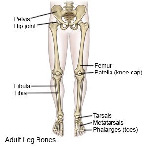
The bones of the leg are the femur, tibia, fibula and patella.the foot bones shown in this diagram are the talus, navicular, cuneiform, cuboid, metatarsals and calcaneus. There are in all 7 bones, which fall under tarsal bones category. This large tendon from the powerful thigh muscles (quadriceps) wraps round the patella and is attached to the top of the lower leg bone (tibia). The tibia and the fibula, at the top of the ankle joint. The hip itself is a ball and socket joint, much like the shoulder.the structures necessary to create this joint are the socket, the joint capsule, muscle, ligaments, and the neck. These are the femur, patella, tibia, fibula, tarsal bones, metatarsal bones, and phalanges (see figure 6.51). The patella (kneecap) is the sesamoid bone in front of the knee. 10 october 2007 (original upload date) source: To explain the term in layman's language, it is the heel bone in the skeletal system. The patella is the kneecap bone. These bones have a marrow, but not a bone marrow cavity. He leg's main function in the human is for use the leg bones diagrams to learn the names of the leg bones and leg anatomy. Some types of leg pain can be traced to problems in your lower spine.
The medial, larger bone of the lower leg. These can include any the following: The bones of the leg and foot form part of the appendicular skeleton that supports the many muscles of the lower limbs. Leg pain can also be caused by blood clots, varicose veins or poor circulation. With different grades of sprains depending on severity.
Leg Bone Diagram Human Anatomy Chart Bones Of The Ankle Leg Bones from i.pinimg.com The bones of the hip include the femur, the ilium, the ischium, and the pubis. The tibia and the fibula, at the top of the ankle joint. Some types of leg pain can be traced to problems in your lower spine. Most of the leg skeleton has bony prominences and margins that can be palpated and some serve as anatomical landmarks that define the extent of the leg. The lower limb contains 30 bones. There are in all 7 bones, which fall under tarsal bones category. Its lower end helps create the knee joint. But the main part of the foot is similar to the hand, with five bones.
Labeled human leg bones created for use in leg bone.
The smaller lateral bone of the lower leg. In addition, the broad hip bones provide protection to the delicate internal organs of the pelvis, such as the intestines, urinary bladder, and uterus. It is where the lower leg bones connect to a large bone in the foot called the talus (say: Related posts of bones leg diagram picture bone anatomy of upper extremity. Disposition of rotator cuff muscles diagram. Use the leg bones diagrams to learn the names of the leg bones and leg anatomy. The thigh bone, or femur, is the large upper leg bone that connects the lower leg bones (knee joint) to the pelvic bone (hip joint). This image is an edited version of this image that was created by user:ladyofhats (mariana ruiz villarreal). These muscles work together to produce movements such as standing, walking, running, and jumping. This allows weight to be distributed either anteriorly or posteriorly throughout the foot. The tibia, commonly known as the 'shin bone', is the largest and most medial of the two.you can palpate its anterior border when you run your finger down the anterior aspect of your leg. These can include any the following: This area is commonly referred to as the calf.
0 Response to "Bones In Leg Diagram - Leg Anatomy / Joints of hand anterior view, lateral view, right hand."


0 Response to "Bones In Leg Diagram - Leg Anatomy / Joints of hand anterior view, lateral view, right hand."
Post a Comment